


PCL Knee Injury, Sprain & Strain
Posterior Cruciate Ligament Injuries
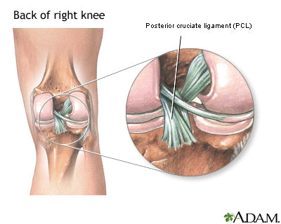
The PCL (also known as the posterior cruciate ligament) is one of the four main ligaments in the knee. Where is your PCL located? The PCL is located in the back of the knee connecting the femur to the shin bone. BraceAbility offers treatment for many knee injuries, including PCL tears. PCL injuries make up less than 20% of injuries to knee ligaments.
The PCL is located in the back of the knee. It is one of several ligaments that connect the thighbone (femur) to the shinbone (tibia). The main function of the PCL is to keep the tibia from moving backward too far and to stabilize the knee. A PCL injury is less common than an ACL injury because the PCL is broader and stronger than the ACL.
BraceAbility offers PCL Knee Braces for a wide variety of injuries to the posterior cruciate ligament including PCL tears, sprains and strains.
See the article “Posterior Cruciate Ligament” for a more detailed explanation of the location of the ligament or see the photo below.
PCL Injury Symptoms
Signs and symptoms of a posterior cruciate ligament injury may include:
- Pain. When the PCL is injured, the pain level is often classified as mild to moderate. As to ACL injuries where the pain is in most cases severe, PCL injuries are not known to cause excessive knee pain unless severe ligament damage takes place.
- Swelling. As is the case with most ligament injuries, if a PCL injury occurs rapid swelling will also occur.
- Instability. Patients who injure their PCL will often describe their knee as feeling “loose”. Another common symptom of a PCL sprain would be the knee giving out. These examples of knee instability are a good sign that a posterior cruciate ligament injury may have occurred.
With most knee injuries, signs and symptoms of the injury are extremely prevalent directly after the injury. This is not the case with many injuries associated with PCL injuries. Signs and symptoms may be so mild that one may not even notice anything is wrong with the knee. The symptoms will continue to get worse, the pain will become more severe and the knee will be less stable if not treated properly.
It is important to see your physician as soon as possible after the injury in order to diagnose the PCL. The initial swelling in the injured knee may be too severe to determine the degree to which the knee is injured. Once the pain and swelling have subsided enough, a sports injury professional should be able to examine the knee.
Most doctors will perform an MRI scan and or an X-Ray to reveal the damage. An MRI is a pain-free procedure that uses radio waves and a strong magnetic field to create computer images of the soft tissues in the body. An MRI scan works very well to determine the degree of injury to ligaments and cartilage because they are soft tissues.
In addition to the MRI, the doctor may also order an X-Ray. An X-Ray cannot detect ligament damage but does reveal if any chunks of bone may have been torn away from the knee in the process. This type of injury is called an Avulsion Fracture.
PCL Sprain & Strain
A PCL sprain is the least common injury associated with sprained knee ligaments. The PCL is the strongest ligament in the knee which makes the likelihood of it being injured the lowest out of the 4 main ligaments. Oftentimes a posterior cruciate ligament injury occurs concurrently with other structures in the knee (cartilage, ACL, MCL, LCL, bone).
When diagnosing a PCL sprain, the patient will receive one of two diagnoses.
Grade 1 Sprain. The ligament is mildly damaged in a Grade 1 Sprain. It has been slightly stretched but is still able to help keep the knee joint stable.
Grade 2 Sprain. A Grade 2 Sprain stretches the ligament to the point where it becomes loose. This is often referred to as a partial tear of the ligament.
Being one of the strongest ligaments in your knee, it is important to understand that a PCL injury requires a powerful force. One of the most common causes of a PCL sprain is a bent knee hitting a dashboard in a car accident or an athlete falling on a knee that is bent. In this injury, the knee is hyper flexed (bent all the way back), with the foot held pointing downwards.
PCL Injury Treatment & Recovery
Directly after the injury takes place, one must apply the principles of R.I.C.E. to the injured knee for 15 minutes every hour to help reduce the pain and swelling as much as possible.
•R – Rest.
•I – Ice.
•C – Compression.
•E – Elevation.
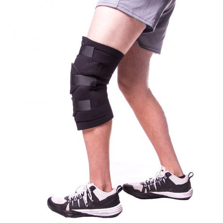
A great option to help reduce pain and swelling while keeping the knee cold is the Knee Wrap with an ice pack. This economical wrap is designed to provide compression and concentrated cold/hot therapy for the injured PCL. This PCL injury brace works like a charm for mild sprains. It’s perfect to wear after sports when suffering from knee pain and helps reduce swelling drastically.
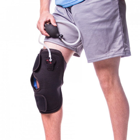
Another great treatment option for an injured PCL is the Inflatable Knee Brace with Cold Therapy. This knee wrap is designed to combine the benefits of pneumatic compression with cold therapy. It features a pump bulb that detaches once desired compression is achieved.
If you find yourself looking for a PCL knee brace to wear while playing sports to protect your sprained PCL, look no further than the Flexion Knee Brace. This PCL injury brace permits full flexion and extension control, which is useful when it comes to meniscus tear treatment and other knee joint problems. The hinged ROM knee brace offers the convenience of an adjustable wrap above the knee while the part below the knee is a sleeve. The CoolTex material that makes up these components make the torn meniscus knee brace ideal for use during non-contact sports and daily living activity.
Check out our full selection of Ligament Knee Braces and Ligament strains and sprains braces.
PCL Rehab and Exercises
Regardless of whether or not you need surgery to repair your injured PCL, rehab is extremely important for all PCL injuries. There are a number of PCL rehab exercises below that will help you get back to 100% in no time.
- Quad sets: Sit on the floor with your injured leg straight and your other leg bent. Press the back of the knee of your injured leg against the floor by tightening the muscles on the top of your thigh. Hold this position 10 seconds. Relax. Do 2 sets of 15.
- Seated quad sets: Sit in a straight-back chair with your injured knee bent at a 90-degree angle. Try to tighten the top of your thigh muscles without moving your leg. Hold for 10 seconds. Do 2 sets of 15.
- Straight leg raises: Lie on your back with your legs straight out in front of you. Bend the knee on your uninjured side and place the foot flat on the floor. Tighten the thigh muscle on your injured side and lift your leg about 8 inches off the floor. Keep your leg straight and your thigh muscle tight. Slowly lower your leg back down to the floor. Do 2 sets of 15.
- Wall squat with a ball: Stand with your back, shoulders, and head against a wall. Look straight ahead. Keep your shoulders relaxed and your feet 3 feet from the wall and shoulder's width apart. Place a soccer or basketball-sized ball behind your back. Keeping your back against the wall, slowly squat down to a 45-degree angle. Your thighs will not yet be parallel to the floor. Hold this position for 10 seconds and then slowly slide back up the wall. Repeat 10 times. Build up to 2 sets of 15.
- Step-up: Stand with the foot of your injured leg on a support 3 to 5 inches high (like a small step or block of wood). Keep your other foot flat on the floor. Shift your weight onto the injured leg on the support. Straighten your injured leg as the other leg comes off the floor. Return to the starting position by bending your injured leg and slowly lowering your uninjured leg back to the floor. Do 2 sets of 15.